History: 35 year old male with knee pain.

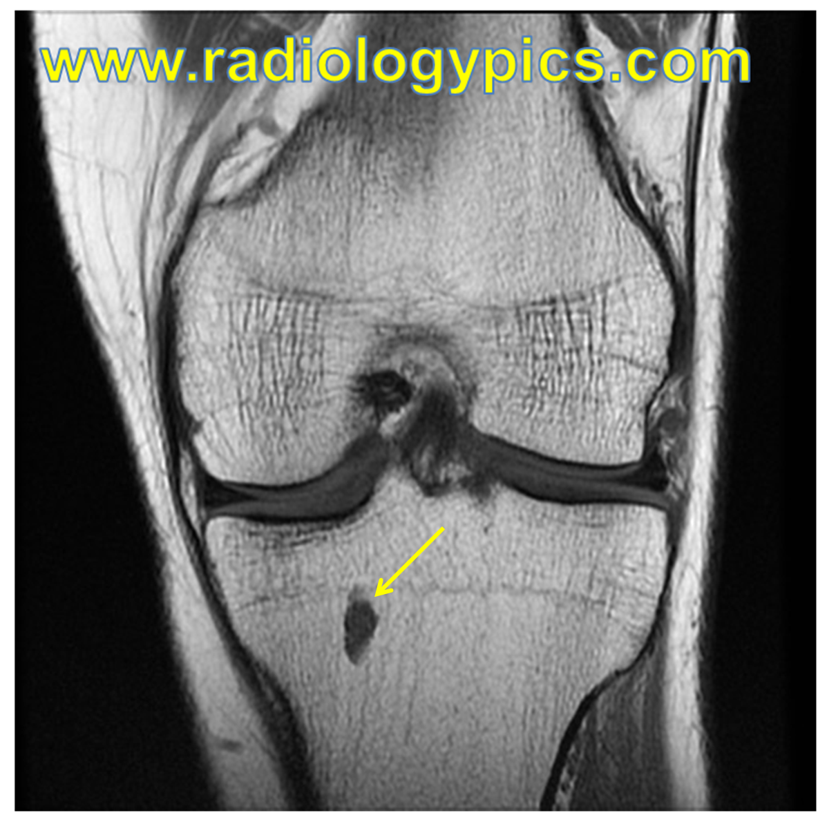
This is the appearance of an enchondroma on MRI. Enchondromas are benign lesions made up of hyaline cartilage that arise within medullary bone. They typically occur in the metaphysis or metadiaphyseal junction of the proximal humerus, proximal and distal femur, and proximal tibia, as in our case above. On MRI, enchondromas are characteristically lobulated with homogeneous hyperintense signal on fluid sensitive sequences, typical of benign cartilaginous lesions.
Ideally, contrast is required to definitively distinguish between enchondromas and low grade chondrosarcomas, as some enchondromas can degenerate into chondrosarcomas. This is shown radiographically by excessive endosteal scalloping, and on MRI with contrast as peripheral puddling of contrast within the lesion. Another distinguishing factor is pain, indicating likely chondrosarcoma. Read more about other distinguishing findings here.
See an enchondroma of a phalanx here.


Leave a reply to Lytic Bone Lesion Mnemonic – FEGNOMASHIC | RADIOLOGYPICS.COM Cancel reply