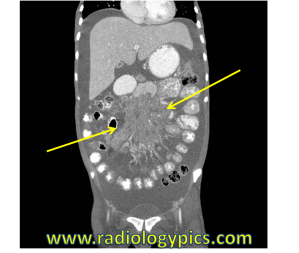
History: 30 year old female with HIV and abdominal pain.


This case demonstrates small bowel mesenteric lymphadenopathy, which is just enlarged lymph nodes in the mesentery. The differential diagnosis along with distinguishing factors is as follows:
1. Lymphoma – the most common malignant cause of mesenteric lymphadenopathy (most commonly Non-Hodgkins Lymphoma). Will commonly show node enlargement in other parts of the body. Treated lymphoma will show calcifications.
2. Cancer/Cancer Metastases – Carcinoid tumors will have a desmoplastic reaction, small bowel tumors, colon cancer, pancreatic cancer
3. Mesenteric Adenitis – usually right lower quadrant, usually children and young adults, classically seen with Yersinia infection
4. Sclerosing mesenteritis – ill-defined fat stranding in the mesentery with lymph node clusters
5. AIDS – Opportunistic infections such as TB or MAC, Kaposi sarcoma, actual HIV direct infection. Kaposi sarcoma will enhance, TB will commonly show necrosis and calcification
6. Other – diverticulitis, ulcerative colitis, Crohn disease, appendicitis, etc.
This case shows a nice example of the “sandwich sign,” which is mesenteric vessels sandwiched between conglomerations of lymph nodes. It is highly suggestive of lymphoma, most commonly Non-Hodgkins Lymphoma.
Thank you to Paul Murphy, M.D. Ph.D. for these images.


Leave a comment