History: 60 year old female with arm pain.
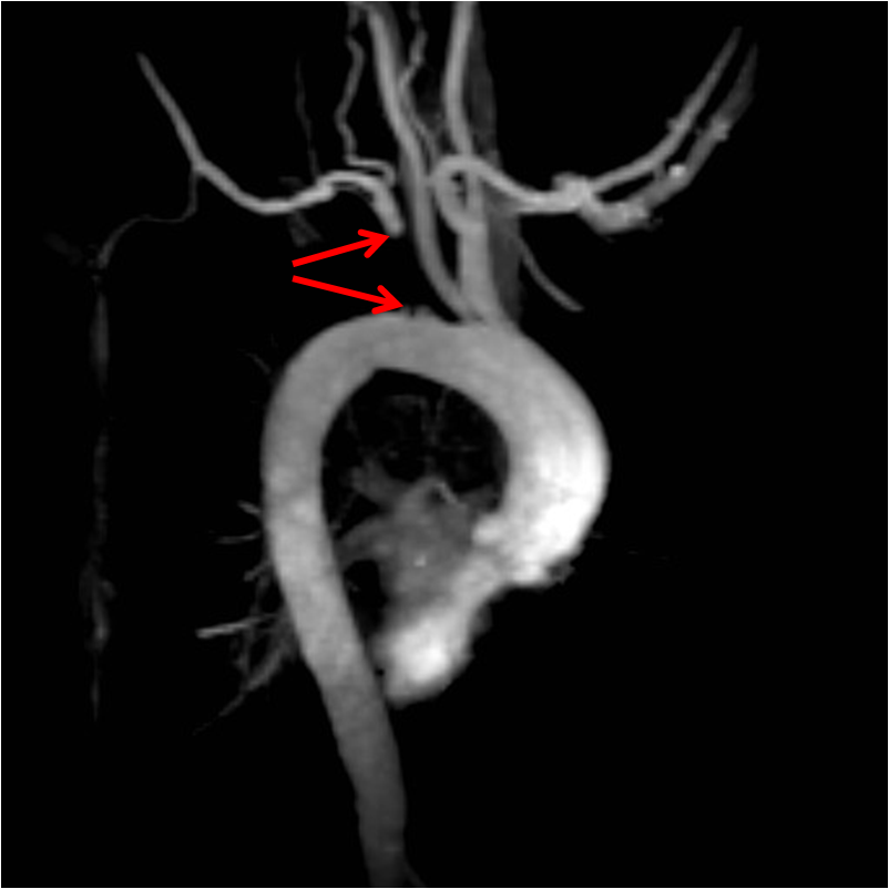
This is a case of subclavian steal syndrome. Subclavian steal syndrome occurs when there is an occluded subclavian artery and collateral blood flow to the distal subclavian artery and arm is provided by reversed flow in the ipsilateral vertebral artery. As seen in the MR angiogram image above, this was due to occlusion of the proximal left subclavian artery. Usually subcalvian steal is asymptomatic, however, when symptoms do occur they usually manifest as exertional arm pain.
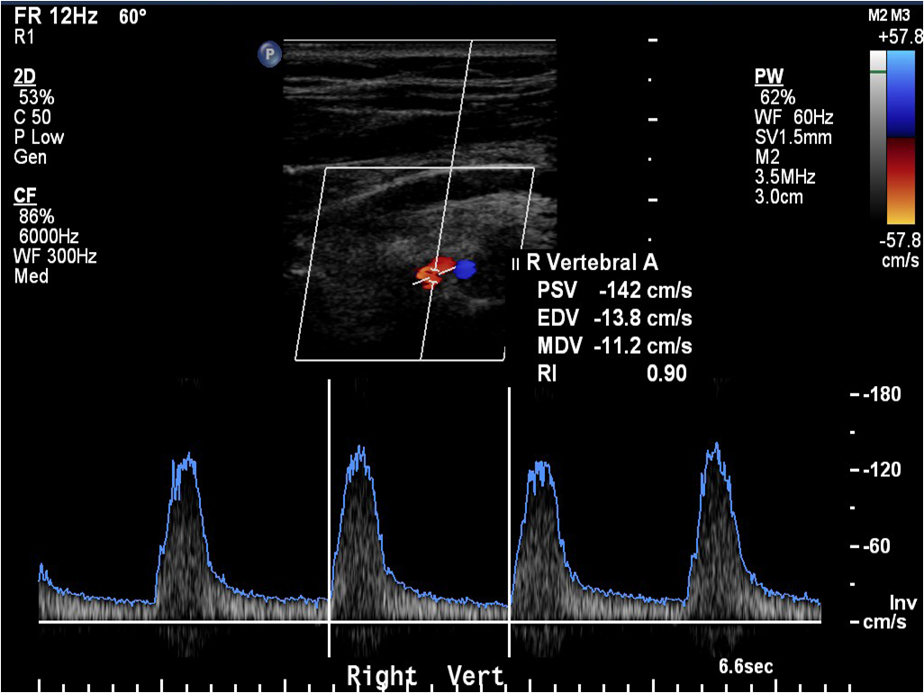
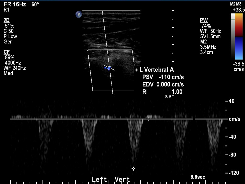
Subclavian steal syndrome is diagnosed on ultrasound by documenting reversal of flow in the vertebral artery, and sometimes with elevated velocity (about >60 cm/sec) in the contralateral vertebral artery. Subclavian steal syndrome can be classified on spectral Doppler ultrasound as mild (slight decreased peak systolic velocity), moderate (alternating biphasic flow, in which provocative maneuvers can cause complete reversal), and complete (no forward systolic velocity towards the head).
It is essential to know the positioning of the transducer and the color scheme chosen by the sonographer, as this can be variable. Traditionally orange/red is towards the transducer and blue is away from the transducer, however as you can see in the images above this can be changed by the sonographer. This is why it is important to look at the directional velocity (the “+” and “-” signs on the scale in the top right of the ultrasound images) and the orientation of the transducer. In the first image above, the transducer is oriented pointing towards the patient’s head, thus flow away from the transducer (in the negative “-” direction) is normal (also note, the spectral waveform can be flipped). In the second ultrasound image above, the transducer is oriented towards the patients feet, and flow is going away from the transducer (in the negative “-” direction again), and this is abnormal. It can get confusing at times!
See an article here about internal thoracic artery doppler in patients with subclavian steal syndrome.
Read about an occluded internal carotid artery here and understand the “string sign” for diagnosing near complete occlusions.
Thanks to Paul Murphy, M.D., Ph.D. for these beautiful images!


Leave a comment