History: 70 year old male with cough.
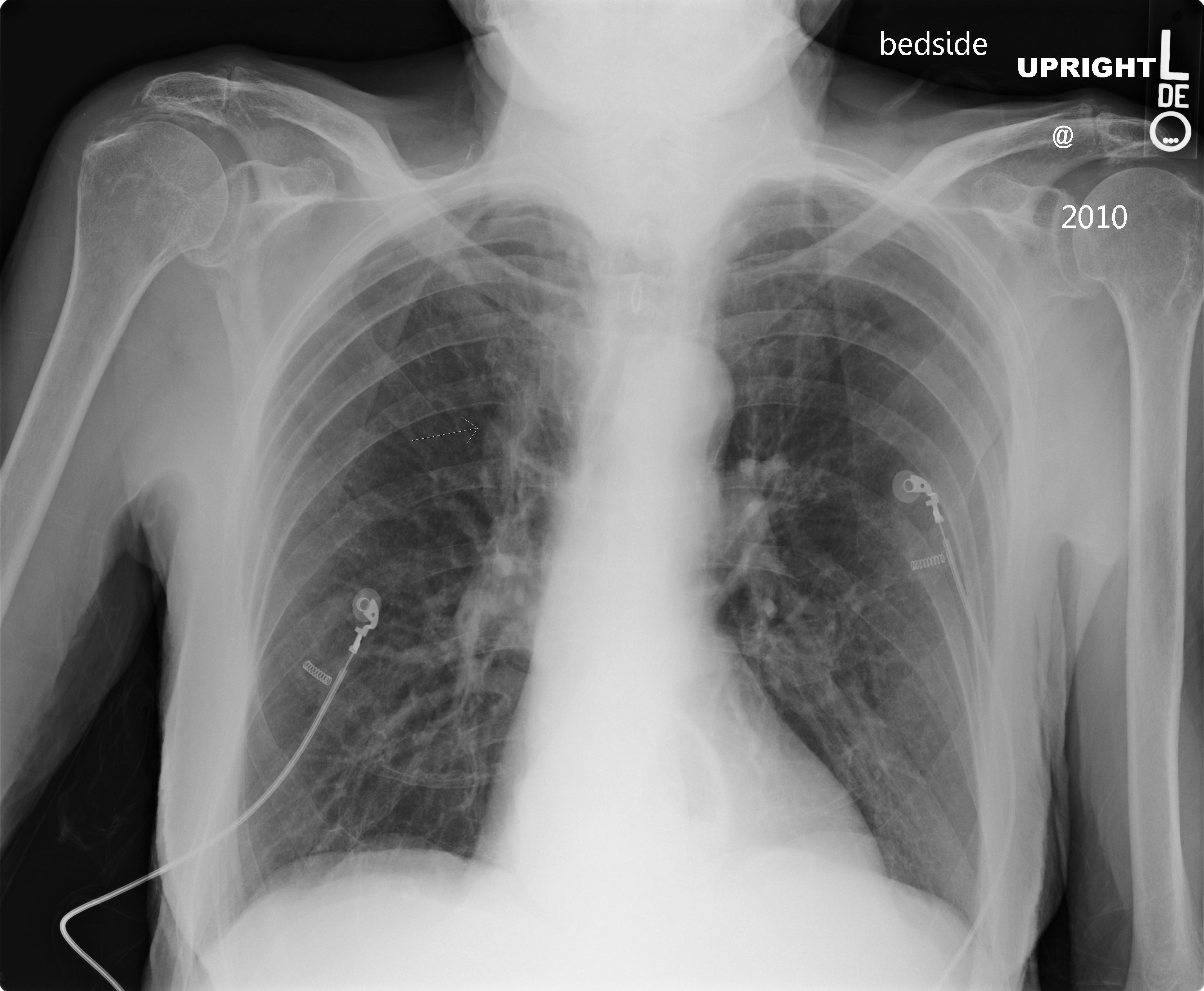

This is a solitary pulmonary nodule (SPN). An solitary pulmonary nodule is a single, focal, rounded opacity that measures less than 3 cm. If larger than 3 cm, it is termed a “mass.” Up to 90% of nodules less than 2 cm are benign. Stability over two years suggests that the process is benign, as well as rapid doubling time of less than 7 days, or prolonged doubling time of greater than 15 months suggest benign entities. Spiculation, as seen in the case above, is more suggestive of malignant processes, while spehrical shape is more suggestive of benign processes. Most lung cancers are solid, but when a solitary pulmonary nodule is seen that is partially solid it has a higher likelihood of malignancy than a solid nodule. The presence of fat suggests hamartoma or lipoma. Malignant processes typically enhance, and up to 90% of them take up FDG on PET studies. An article regarding the morphologic evaluation of SPNs is found here.
The differential diagnosis for a solitary pulmonary nodule includes granuloma, lung cancer, intrapulmonary lymph node, hamartoma, carcinoid, solitary metastasis, infectious/inflammatory process, arteriovenous malformation, and others mimics such as nipple shadows (on radiographs), and pulmonary venous confluences.


Leave a reply to Pneumonia | RADIOLOGYPICS.COM Cancel reply