History: 50 year old male with productive cough.

Frontal radiograph of the chest reveals tubular opacities in the lung bases and right mid lung.
Frontal radiograph of the chest reveals tubular opacities in the medial lung bases and right mid lung.
CT of the thorax in the same patient reveals multiple areas of bronchiectases filled with mucus (arrrows), with a more focal area of consolidation in the right middle lobe (double arrows).
CT of the thorax in the same patient reveals multiple areas of cylindrical bronchiectases filled with mucus (arrrows), with a more focal area of consolidation in the right middle lobe (double arrows).

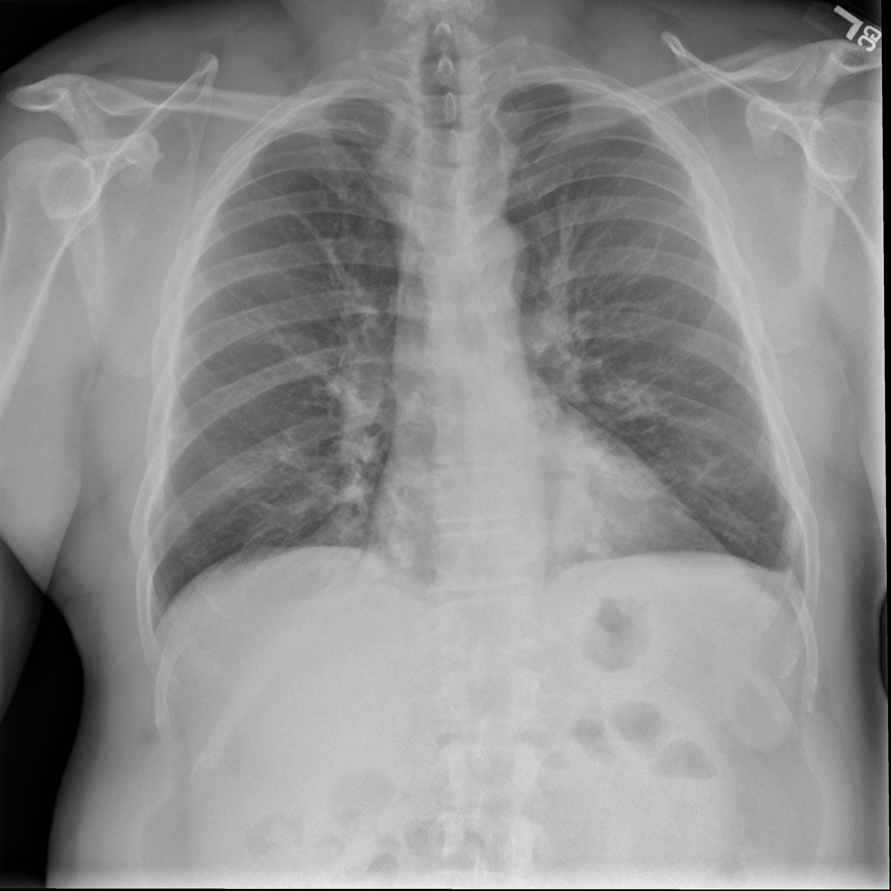
This is a case of allergic bronchopulmonary aspergillosis (ABPA). ABPA is a hypersensitivity reaction (Type 1, mediated by IgE and IgG) most commonly to aspergillus fumigatus. It commonly occurs in patients with cystic fibrosis. The round opacities in the CT image above represent dilated bronchi impacted with mucus, and have been called the “finger in glove” sign. The differential diagnosis includes bronchial obstruction with bronchogenic carcinoma, bronchial atresia, foreign body bronchial obstruction, and bronchocentric granulomatosis.

One response to “Allergic Bronchopulmonary Aspergillosis”

  1. […] foreign bodies, traction bronchiectasis seen in interstitial lung disease, cystic fibrosis, asthma, allergic bronchopulmonary aspergillosis, and immotile cilia syndrome (Kartagener […]

Leave a reply to Bronchiectasis – Differential Diagnosis | RadiologyPics.com Cancel reply

Trending