History: 60 year old male with left sided chest pain.

Coronal contrast enhanced CT of the thorax shows a large lobulated soft tissue mass within the left lower hemithorax which invades the diaphragm to enter the abdomen. Also note the large soft tissue mass which invades the left lateral fifth rib.
Maximum intensity projection (MIP) of an F18 FDG-PET/CT scan shows the large left hemithorax mass which has marked FDG uptake. Multiple other lesions with avid uptake are seen, concerning for metastatic disease.

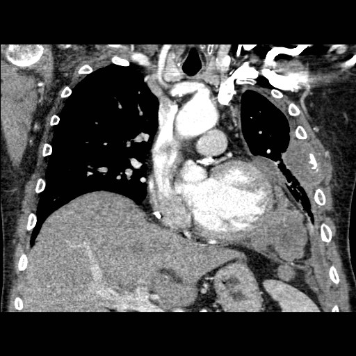
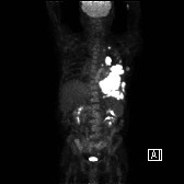
This is a case of pathology proven metastatic malignant mesothelioma. These are tumors of the pleura, and are most likely secondary to long-term asbestos exposure, in which there is a dose-response relationship and typically a latent period of 30-45 years. Asbestos plaques are not a precursor to malignant mesothelioma. While the aggressiveness of this particularly mass makes malignant mesothelioma the likely diagnosis, the differential includes metastatic adenocarcinoma, empyema, malignant thymoma, splenosis, and lymphoma.

6 responses to “Metastatic Malignant Mesothelioma”

  1. […] diagnosis for the right lateral chest wall mass visualized in the three MR images above includes mesothelioma, primary lung carcinoma, soft tissue sarcomas such as fibrosarcoma and malignant fibrohistiocytoma, […]

  2. Good day! This is kind of off topic but I need some guidance from an established blog. Is it tough to set up your own blog? I’m not very techincal but I can figure things out pretty fast. I’m thinking about creating my own but I’m not sure where to begin. Do you have any tips or suggestions? Thanks

  3. Its very easy. Just go to wordpress.com and peruse the help section and go from there.

  4. […] pneumonectomy, community acquired pneumonia, bronchogenic carcinoma, pleural masses such as mesothelioma, and finally pulmonary […]

  5. […] extrapleural fat. Pleural malignancies are an additional consideration, as plaques can be seen with mesothelioma and pleural metastases. Finally, pleurodesis can cause this […]

  6. […] – Mesothelioma  A – Adenocarcinoma L – Lymphoma L – Leukemia E – Empyema T – Thymoma […]

Leave a reply to news article Cancel reply

Trending