History: 60 year old male lost to follow up now returns five years later for reevaluation of a known right renal mass.
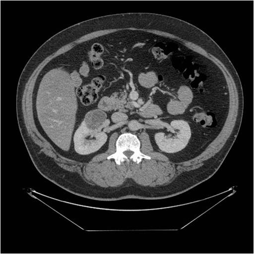
This mass is classified as a Bosniak 3 lesion given the enhancing internal septations and enhancing capsule; therefore, it is required to be surgically removed for definitive differentiation between malignancy and multilocular cystic nephroma. Given that it was stable for 5 years when compared to the baseline imaging, multilocular cystic nephroma was favored, which was the final diagnosis on pathology.


Leave a reply to Solution to Unknown Case #24 – Multilocular Cystic Nephroma on Ultrasound | RADIOLOGYPICS.COM Cancel reply