History: 35 year old male with foot pain. 
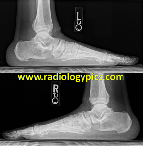
Pes Planus - Lateral radiographs of both feet show marked flattening of the longitudinal arch and overlap of the metatarsals, compatible with pes planus and pronation/valgus of the forefoot.
Pes Planus – Lateral radiographs of both feet show marked flattening of the longitudinal arch, increased talocalcaneal angle and overlap of the metatarsals, compatible with pes planus and pronation/valgus of the forefoot.

This is the appearance of pes planus, or flatfoot, which occurs in up to 20% of adults with no other abnormalities.

There are many contributors to pes planus deformity, including hindfoot valgus shown by increased talocalcaneal angle to greater than 45 degrees, midfoot sag or lisfranc ligament injury, and forefoot pronation (seen as overlapping metatarsals in the images above).  Pes planus can be idiopathic, due to posterior tibialis tendon injury, charcot joint, lisfranc ligament injury, rheumatoid arthritis, or tarsal coalition. It is important to always obtain weightbearing films to assess for alignment, and compare with the contralateral foot.

Flatfoot in a 55 years old female with ankle a...
Flatfoot in a 55 year old female with ankle and knee arthritis (Photo credit: Wikipedia)

One response to “Pes Planus”

  1. […] Pes Planus – RADIOLOGYPICS.COM | … – Pes Planus – Lateral radiographs of both feet show marked flattening of the longitudinal arch, increased talocalcaneal angle and overlap of the metatarsals …… […]

Leave a comment

Trending