
History: 1 day old infant with bilious emesis.

This is a normal upper GI study in a neonate. The patient is usually imaged first in the right lateral position and multiple images are obtained as contrast passes through to the duodenum and ligament of Treitz. The most common indication in the emergent setting is bilious emesis, rule out malrotation. The lateral image is below.
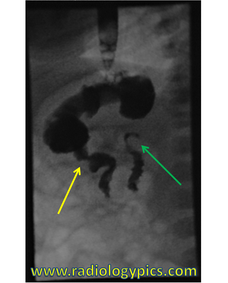
After the lateral images are obtained, usually just one to two images in the frontal projection are sufficient to demonstrate the anatomy. The radiologist should look for abnormal location of small bowel loops, or even twisting of small bowel loops, called midgut volvulus.


Leave a comment