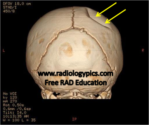
History: Infant status post fall from 4 feet.

This is the appearance of a depressed skull fracture in an infant. In the setting of skull fractures in an infant, one always has to rule out the presence of non-accidental trauma (NAT), which is medical-speak for child abuse. There are multiple characteristics of skull fractures which lead one to believe it was the result of an accident, such as a short segment linear fracture through the parietal bone. Features that suggest non-accidental trauma (read: “child abuse”) include multiple or complex fractures, large fractures, depressed fractures (seen above), more than one bone, non-parietal bone involvement, and intracranial hemorrhage (read about non-traumatic intracranial hemorrhage here).
Given the finding of depressed skull fracture in this case, we have to be worried about non-accidental trauma; however, we also always have to evaluate the whole picture and order a social work consult. In this case, the social worker and clinician came to the conclusion that this actually was accidental trauma, which makes for a nice happy ending.


Leave a comment