History: 10 year old male status post fall.
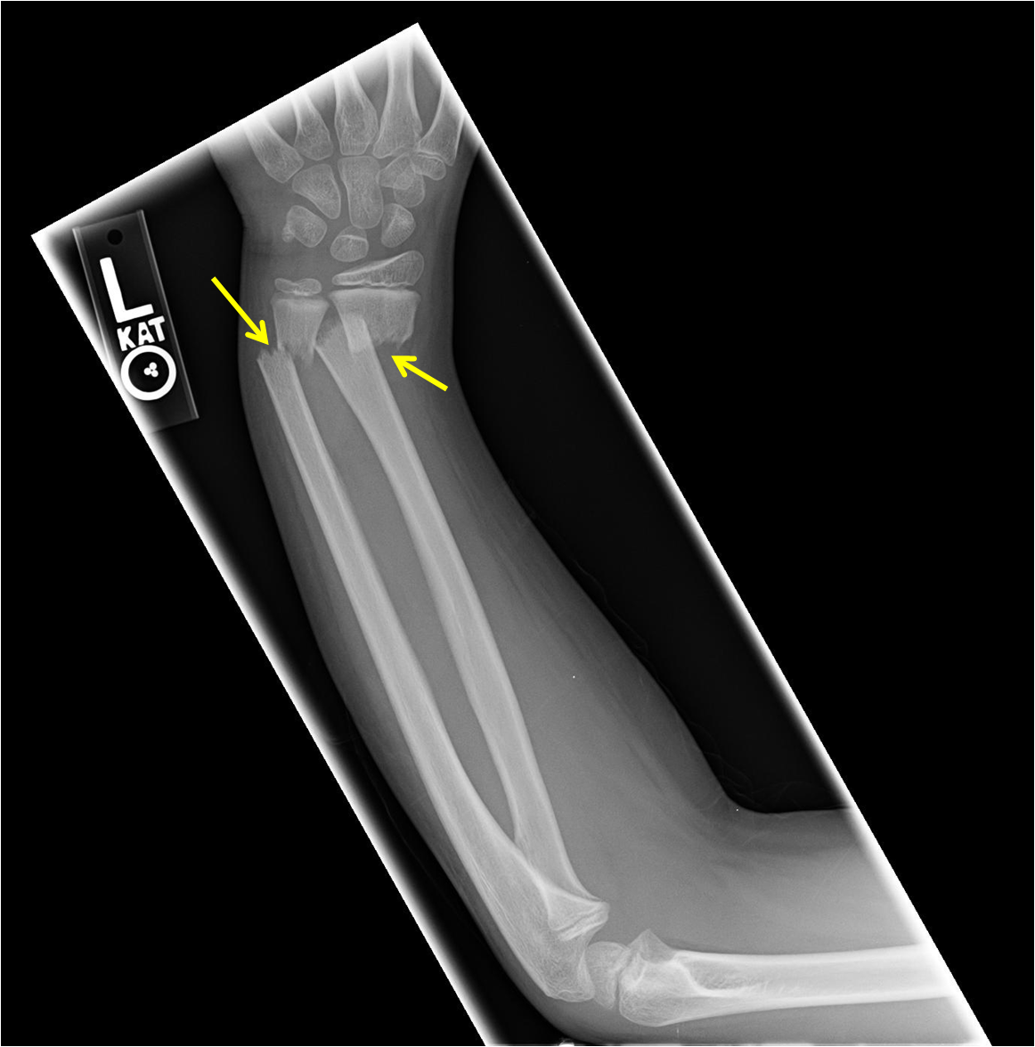
This is the appearance of a both bone forearm fracture in the pediatric patient. The most common mechanism is fall on an outstretched hand, sometimes referred to as FOOSH; however, this mechanism of injury typically causes other types of distal forearm fractures such as a Colles fracture or Smith fracture.
Most of these fractures are treated without surgery via closed reduction and immobilization for 6-12 weeks.
As a side note, this is obviously a skeletal immature patient. How can you tell the age? While there is a wide degree of variation, the carpal bone ossification pattern indicates the patient is likely near 11 years old, as an ossified pisiform is seen.


Leave a comment