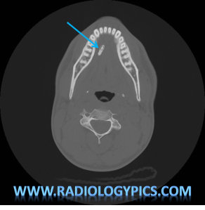
History: 35 year old male with lump underneath tongue.

This is a pretty picture of a sialolith, which essentially is a calcified stone in a salivary gland duct. They most commonly form in the submandibular gland duct (Wharton’s duct) due to its long and upward going course. Sialolithiasis presents with pain and swelling of the involved gland, often every time the patient eats or even thinks about eating, as this stimulates salivation.
A case from the New England Journal of medicine shows pictures of a sialolith, similar in size to the case above.


Leave a comment