History: 50 year old man with jaw pain. 


Coronal CT image of the face reveals a periapical lucency near one of the right maxillary molars (yellow arrow). Note the mucosal thickening in the right maxillary sinus as a result of the adjacent inflammation.
Periapical tooth abscess: Coronal CT image of the face reveals a periapical lucency near one of the right maxillary molars (yellow arrow). Note the mucosal thickening in the right maxillary sinus (green arrow) as a result of the adjacent inflammation.

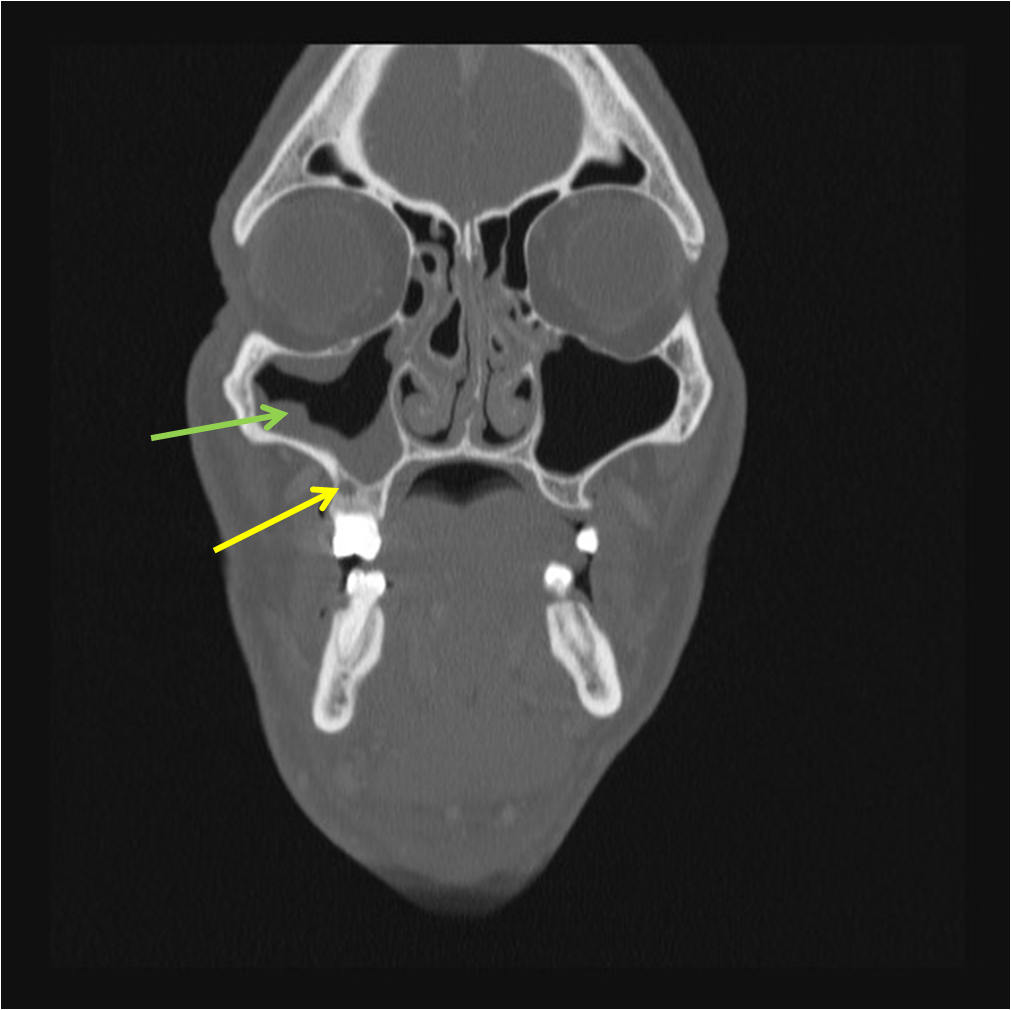
This is an example of a tooth abscess, or periapical abscess. The lucency in the maxilla indicated by the yellow arrow is somewhat nonspecific and may be secondary to tooth loosening, however, the adjacent maxillary sinus mucosal thickening argues for the case of an abscess. Click the following link to learn about Odontogenic Sinusitis.


The spectrum of apical periodontitis (inflammation at the root of the tooth, beginning in the periodontal ligament) includes periapical granuloma, periapical abscess, and periapical radicular cyst. Another term used is rarefying osteitis, which is loss of bone due to adjacent inflammation. Rarefying osteitis is the radiological term to describe the clinical entity of apical periodontitis. Treatment includes tooth extraction.


Tooth #5, the upper right first premolar, afte...
Tooth #5, the upper right first premolar, after extraction. The two single-headed arrows point to the CEJ, which is the line separating the crown (in this case, heavily decayed) and the roots. The double headed arrow (bottom right) shows the extent of the abscess that surrounds the apex of the palatal root. (Photo credit: Wikipedia)

One response to “Periapical Tooth Abscess – The spectrum of apical periodontitis”

  1. […] tooth, whether there is a dental carie, or in this case, a periapical lucency suggestive of a periapical abscess. It is treated with tooth extraction and […]

Leave a comment

Trending