Superficial Venous Thrombosis to Deep Venous Thrombosis
History: 55 year old female seen five days earlier with a diagnosis of superficial thrombophlebitis returns with continued and worsening leg swelling. This is a case of extension of a superficial venous thrombus into the deep venous system of the leg from the short saphenous vein to the popliteal vein. In patients with superficial…
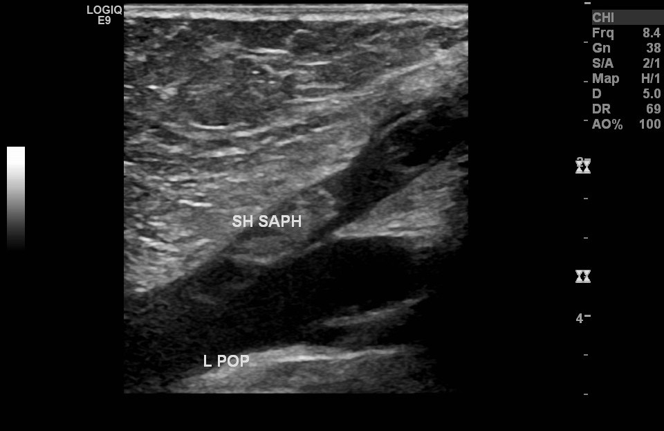
History: 55 year old female seen five days earlier with a diagnosis of superficial thrombophlebitis returns with continued and worsening leg swelling.
Superficial Venous Thrombus: Transverse ultrasound image through the left calf with and without compression technique shows a distended, noncompressible short saphenous vein containing echogenic clot material. The popliteal vein is compressible at this level.Superficial Venous Thrombus: Sagittal ultrasound of the left calf in the same patient at the junction of the short saphenous and popliteal vein again shows completely occlusive clot with no flow in the short saphenous vein. Note, the popliteal vein is patent.Superficial Venous Thrombus extending to Deep Venous Thrombus: Sagittal ultrasound at the same location as the image above without doppler now shows clot material which extends to the junction of the short saphenous and popliteal vein, indicating extension of superficial thrombus into the deep venous system of the left leg.
This is a case of extension of a superficial venous thrombus into the deep venous system of the leg from the short saphenous vein to the popliteal vein. In patients with superficial venous thrombosis, 25% will have coexisting deep venous thrombosis or pulmonary embolism and up to 10% will have future extension of the superficial thrombus into the deep venous system. This patient should be treated with anticoagulation. Other potential complications are listed here.
[…] This is a case of acute pulmonary embolism. A pulmonary embolism is a blood clot which travels via the heart into the arteries that supply blood to the lungs. Typically the blood clot arises from the lower extremities, what is known as a deep vein thrombosis. […]


Leave a comment