History: 18 year old female with pelvic pain.

Transverse ultrasound image through the pelvis shows two separate widely splayed uterine horns, which may be seen in both uterine didelphys and bicornuate uterus.
Uterine Didelphys: Transverse ultrasound image through the pelvis shows two separate widely splayed uterine horns, which may be seen in both uterine didelphys and bicornuate uterus.
Transverse ultrasound through the pelvis in the same patient at a more inferior section shows two separate cervices, defining this abnormality as a uterine didelphys.
Uterine Didelphys: Transverse ultrasound through the pelvis in the same patient at a more inferior section shows two separate cervices, defining this abnormality as a uterine didelphys.

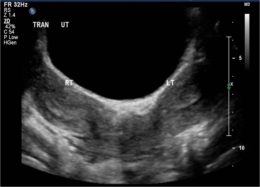
This is the classic appearance of uterine didelphys. Mullerian duct anomalies are classified according to the American Fertility Society system here. Occasionally a uterine horn can become obstructed and cause hematocolpos, or hematometrocolpos.

One response to “Mullerian Duct Anomalies: Uterine Didelphys”

Leave a comment

Trending