History: 65 year old male with a history of chronic lymphocytic leukemia presents with left sided chest pain, shortness of breath, and hypoxia after a CT-guided biopsy of a left lung mass.
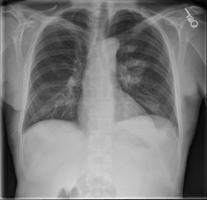
Pneumothorax: Single frontal upright chest radiograph demonstrating a moderate sized left pneumothorax at the left lung apex. A left hilar cavitating ground glass opacity and an additional anterior left upper lobe ground glass opacity are seen.
This is an iatrogenic pneumothorax in a patient status post lung biopsy. This patient had a cavitary lung mass.
Discussion
Trackbacks/Pingbacks
Pingback: Penetrating Chest Trauma – Knife in Chest | RadiologyPics.com - February 20, 2013
Pingback: Unilateral Hyperlucent Hemithorax Mnemonic – CRAWLS | RADIOLOGYPICS.COM - March 16, 2013
Pingback: The ABCs of Trauma Mnemonic – ABCDEFGHI | RADIOLOGYPICS.COM - March 19, 2013
Pingback: Solution to Unknown Case #23 – Empyema (loculated pleural effusion) | RADIOLOGYPICS.COM - March 21, 2013